NMR Sample Preparation
Sample Transfer into NMR Tubes
Although our lab seldom produces NMR samples we frequently have to transfer the samples to an NMR tube. According to different requirements, three kinds of NMR tubes are generally used in the Szyperski Lab: regular 5 mm NMR tubes, Shigemi tubes and capillary tubes. Each kind of tube has its own special sample preparation procedure.
Regular and Shigemi Tubes
Regular NMR tubes:
- Pros
- Easy to transfer or titrate sample.
- Low price.
- Durable.
- Can be used with any spectrometer brand.
- Cons
- Requires large sample volumes - 0.4 to 0.6 ml.
- Suboptimal shimming, consequently, water suppression and sensitivity are poor (and smaller the volume, worse is the effect).
- Sample is subject to air oxidation.
Shigemi NMR tubes:
- Pros
- Ideal for small sample volumes: 0.2 to 0.3 ml
- Optimal shimming and water suppression
- Better sample stability due to reduced air oxidation
- Cons
- Complicated transfer and handling.
- High price.
- Very brittle - easy to break.
- Different tube types for different spectrometer brands.
In general, it is preferable to use Shigemi tubes for the [100% 13C, 100% 15N]-labeled samples. For [5% 13C, 100% 15N]-labeled samples, regular tubes are also fine, since these samples are used to record in general the 13C-HSQC spectrum of the methyl region, where water suppression is not an issue.
NOTE : It is advised to use long-end pipettes to transfer sample solution into and out of regular 5 mm NMR tubes and Shigemi tubes.
Protocol to transfer sample in a regular tube:
Fig.1 Sample in regular NMR tubes
- Place the sample at the bottom of the tube and cap the tube.
- Spin the tube in the hand centrifuge to collect residual sample from the walls and colapse the air pockets and bubbles, if any. Wrap the tube in a tissue for padding before inserting it into a holder of the centrifuge.
- If a part of the sample collected in the cap, transfer it into the tube and spin the sample again.
- Wrap a thin strip of Parafilm around the seam between the tube and its cap to prevent the sample from drying out.
- Put a label on the tube.
Protocol to transfer sample in a Shigemi tubes:
Fig.2 Sample in Shigemi tubes
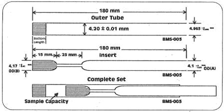
The Shigemi tube is composed of two parts: the outer tube and the insert (also called plunger). The insert is made of a special type of glass with magnetic susceptibility matching to that of the solvent (H2O for protein NMR). The matching magnetic susceptibility removes the edge effect at the interface of the sample and the glass, thereby improving the shimming. The whole set is expensive (~ $80 per set) and the glass type is more brittle as compared to the regular tubes. Extra care should therefore be taken while handling Shigemi tubes.
- Place the sample at the bottom of the tube and cap the tube.
- Spin it in the hand centrifuge to collect residual sample from the walls and collapse the air pockets and bubbles, if any. Wrap the tube in a tissue for padding before inserting it into a holder of the centrifuge.
- If a part of the sample collected in the cap, transfer it into the tube and spin the sample again.
- Slowly push the insert down the tube to drive the air out.
- At this point, it is common to get bubbles under the insert. To get rid of them, hold the tube at an angle (and maybe knock on it a few times) - the bubbles should then settle at the interface between the insert bottom and the tube wall. Push forward quickly, but gently, and simultaneously rotate the insert (I have never tried rotating - sounds a bit complicated. Does it work better this way? Main.AlexEletski).
- If you have a lot of bubbles (or foam) in the reservoir then let the sample stand still for some time. After a while the bubbles will rise on their own. Keep the inset in place by wrapping a thin strip of Parafilm around the tube rim.
- Pull the insert out as far as possible without letting any air back in.
- Repeat steps 5 - 7. if necessary.
- Wrap a thin strip of Parafilm around the tube rim. This should keep the insert in place as well as prevent the sample from drying out.
- Label the tube
Key notes:
- Rotate the insert when moving it in or out to reduce the resistance.
- Be careful not to drive the insert too far, especially when pushing air bubbles out. The neck of the insert forms a reservoir in the tube, but once it is full the sample will leak out.
- When immersed in liquid, the insert may easily slide under it own gravity. Therefore, always fix the insert with Parafilm when leaving the sample unattended
- It is important to push air bubbles out. Even the tiniest bubbles can lead to bad shimming and broad water lines - and that is exactly what Shigemi tubes are designed to avoid.
- Waiting for foamy solution to settle down before pulling up the insert can be crucial for maximizing the sample height. With certain viscous solutions, such as phage-based alignment media, the time required may be as long as several hours.
Maximum Volume
Varian tubes (BMS-005TV) have bottom length of 15-16 mm, leaving 16 mm as the maximul sample depth. This corresponds to 260 ul at 4.52 mm inner diameter. You still need to have some liquid filling part of the reservoir, meaning that you probably have to put 280-300 ul into the tube.
It is possible to use Bruker shigemi tubes with bottom length of 8 mm in Varian spectrometers. In this case max sample depth would be 24 mm, corresponding to 385 ul.
Capillary Tubes
We use 1.0mm and 1.7mm O.D. tubes from Wilmad. The capillary action (with a little help) is always sufficient to draw the solution into the tube. Normally, aliquot ~30uL of solution into a small Eppendorf tube, dip the capillary in, and tip or invert the whole setup if the solution does not completely go into the capillary. If needed, you can apply some suction from a syringe or pipette or even 'inject' the solution into the capillary with a syringe and very thin needle although these methods have a higher risk of squirting your solution onto the bench.
When sealing the tubes, first start with a good length of capillary tube (almost as long as the NMR tube) and put sample solution in it. Then tip the capillary until the solution moves to the middle of the tube. Now it is safe to seal the end of the tube in a Bunsen burner flame -- place only the very tip of the tube into the flame. To avoid the possibility of the protein heat-denaturing while sealing in case of insufficient space at the end of tube, wrapping the tube with some form of a 'heat-sink' (like a wet towel placed in a freezer) can be tried. After sealing one end of the tube, centrifuge the whole set-up (place sealed end into the centrifuge) by using a small hand-operated centrifuge for a few seconds to spin down the solution into the sealed end of the capillary tube. Seal the other end of the tube. As the second end begins to seal, essentially a closed container is heated. Normally, bubbles will generate on one end of the tube.
Some tips for handling capillary tubes and lowering the sample temperature:
- Don't use capillaries that have been broken on both ends (i.e., keep at least one machined end of glass to prevent particles of glass getting into your solution)
- Carefully clean and dry all of the capillaries
- Try to keep the tubes perfectly parallel to the magnetic field by using empty tubes or small pieces of paper wrapped around the top of the capillary tube to keep everything aligned and motionless in the NMR tube
- Try a few times with H2O/D2O and see how it goes
- Before you start, centrifuge the solution at ~14000g for ~30minutes
- Use a 1D (no water suppression) and array the temp from -5C to -20C in -0.1C steps to monitor the cooling. (if one out of ten capillaries freeze, then a decrease in the intensity of the water line could be observed)
- Don't spin the sample or bump the spectrometer during the cooling progress.
Short-term and Long-term NMR Sample Storage
All NMR samples should be labeled and stored at 4C or -20C. The label should include the sample name and concentration, all buffer components, percentage of H2O:D2O, initials of the preparer and the date prepared. This should be neatly printed of prepared with a word processing program. You may also consider a code that is cross referenced to your lab journal giving more detailed information about the sample preparation and the sample conditions.
The optimal method for short-term sample storage, provided the sample is stable and sodium azide is present, is to simply keep them at 4C. There are plenty of 10 mm tube racks in the refrigerator. For valuable samples you should place the NMR tubes inside a second container, e.g., a 10mm capped NMR tube or a homemade container that will serve to catch any spilled sample in the event of glass breakage. This doubly protected NMR sample tube should then be placed in a 10 mm rack.
The optimal method for long-term sample storage, provided they are stable upon freezing, is to transfer the solution to a 1.5 mL plastic Eppendorf tube and then freeze at -20C. The sample can also be lyophilized at this stage. Lyophilized protein (r.t. or -20C) is probably the best condition to store biological sample for extended periods of time.
Precautions: DO NOT freeze samples directly in the glass NMR tubes, particularly in expensive Shigemi tubes. The risk of glass breakage and sample loss is high. If you need to freeze an NMR sample directly in the tube, then the following precautions must be taken: The liquid should be spread uniformly on the glass surface prior to freezing in order to prevent breaking of the thin glass walls; The NMR tube containing a frozen sample should be placed in a clean container, e.g., a 10 mm capped NMR tube or a homemade container that will serve to catch any spilled sample in the event of glass breakage; The sample must be stored in a 10 mm rack provided for this purpose.
-- Main.Gaohua.Liu - 11 Feb 2007

